Friday, 20 January 2017

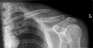
The Rising Incidence of Operative Treatment of Mid-shaft Clavicle Fractures

State Emergency Department Databases (SEDD) and State Ambulatory Surgery Databases (SASD) were used to identify patients presenting with mid-shaft clavicle fractures from 2005 to 2010 in California and New York State. Patients were identified by International Classification of Disease, Ninth Edition (ICD-9) and Current Procedural Terminology (CPT) codes. Multivariable logistic regression analysis was conducted to illustrate any demographic trends regarding patients undergoing operative fixation.

Mid-shaft Clavicle Fractures
Results: Operative fixation of mid-shaft clavicle fractures increased by 368% and by 349% in California and New York, respectively, while the number of patients with clavicle fractures presenting to emergency departments remained stable.

Conclusion: The incidence of operative fixation of mid-shaft clavicle fractures has increased substantially at a similar rate in two states over a short period of time.
PDF LINK
Thursday, 19 January 2017

Intra-articular Injections for Osteoarthritis

The two articles in this month’s press of The Journal of Arthritis, one by Ekinci et al. titled ‘A New Treatment Option in Osteoarthritis: Prolotherapy Injections’ and the other by Senatorov et al. titled ‘Clinical Outcome of Hylan G-F 20 Injections in Shoulder and Hip.

Osteoarthritis
Osteoarthritis: A Retrospective Review’ raise several important points surrounding the topic of intra-articular injections. Firstly that there is a paucity of high quality literature in the field that often leads to inconclusive or misrepresented conclusions on drug efficacy in established treatments. The articles also highlight the heterogeneity in injection procedure, site, concentration, preparation and outcome measurements.

This editorial will, therefore, provide a concise review on some of the current injection therapies and suggest possible improvements to methodological structure to inspire readers strive for high quality data.

Wednesday, 18 January 2017

Plantar Fasciitis: What Does the Evidence Show?

Plantar Fasciitis
Plantar Fasciitis (PF) is caused by repetitive and excessive tensile stress applied to the PF, which leads to fibrosis and inflammatory and degenerative changes. The PF is a membrane that connects the heel bone and the toes, and has a thick structure with bonding strength. PF plays a complex role in the protection and support of the foot itself, and is also important for support of body weight.

There are several well-characterized causes of plantar fasciitis: A flat foot, a supinated foot, a tight Achilles tendon, age, excessive walking or standing, poorly cushioned footwear, obesity, occupations with prolonged standing, pes planus (flat feet), pes cavus (high-arched feet), overpronation.

weak plantar flexor and intrinsic muscles of the foot, running for exercise or competition and other conditions that reduce the shock-absorbing power of the feet. PF is painful, can alter daily activities and presents as a sharp pain localized to the plantar foot and medial heel.

Tuesday, 17 January 2017

Crystal Induced Arthropathies

 Arthropathies
Acute gout most frequently involves the first metatarsophalangeal (MTP) joint at first presentation. However, it has non-specific radiographic features. Chronic gout has a characteristic radiographic appearance with tophus considered to be a hallmark feature.

Tophi are seen as focal eccentric soft tissue swelling of the affected joints or soft tissues resulting in a "lumpy bumpy appearance" and cause well-defined punched out juxta-articular erosions with sclerotic rims and overhanging edges due to pressure on the adjoining bone. Normal bone mineralisation and joint spaces are preserved till late in the disease.

It occurs in asymmetrical polyarticular distribution with involvement of the feet, ankles, knees, hands, and elbows, in decreasing order of frequency

Monday, 16 January 2017

Intra-articular Injections for Osteoarthritis: From Bench to Bedside, can we Teach Old Drugs New Tricks?

Osteoarthritis
The two articles in this month’s press of The Journal of Arthritis, one by Ekinci et al. titled ‘A New Treatment Option in Osteoarthritis: Prolotherapy Injections’ and the other by Senatorov et al. titled ‘Clinical Outcome of Hylan G-F 20 Injections in Shoulder and Hip Osteoarthritis: A Retrospective Review’ raise several important points surrounding the topic of intra-articular injections.

Firstly that there is a paucity of high quality literature in the field that often leads to inconclusive or misrepresented conclusions on drug efficacy in established treatments. The articles also highlight the heterogeneity in injection procedure, site, concentration, preparation and outcome measurements.


This editorial will, therefore, provide a concise review on some of the current injection therapies and suggest possible improvements to methodological structure to inspire readers strive for high quality data. Further, through an example of a possible novel therapy, Actovegin, this editorial will hopefully challenge readers’ way of thinking to consider the reapplication of established drugs; posing the question can you teach an old drug new tricks?

Thursday, 12 January 2017

Antioxidant Enzymes in Rheumatoid Arthritis

Joint destruction in rheumatoid arthritis (RA) is due to tissue injury in the area caused by inflammatory reactions, release of MMPs and free radicals produced by neutrophils and macrophages.

Rheumatoid Arthritis
The control of free radical production may have therapeutic roles thus the study was done to check the status of lipid peroxidation product malondialdehyde (MDA) and a few antioxidant enzymes in RA patients.

45 RA patients and 40 controls were selected. Controls were asymptomatic and RA patients were selected according to ACR criteria. RA patients had significantly high MDA, SOD and ALP and reduced activity of catalase and GR as compared to controls. SOD showed positive correlation with ALP.

Tuesday, 10 January 2017

Systemic Amyloidosis with Predominant Spine Involvement

Primary systemic amyloidosis with main bone involvement is a rare disease. Clinical symptoms and radiographic findings are usually nonspecific and may be confused with primary bone tumor, metastatic disease, metabolic disorders or infections. The occurrence of an amyloidoma in the spine is rare.

Predominant Spine Involvement
We describe the case of a 57-year old man presenting with a pathological fracture of T7 in presence of a large, soft tissue mass narrowing the spinal canal, responsible for a worsening paraplegia.

Diagnosis of systemic amyloidosis was made after surgical treatment. Pathological examination showed an amorphous eosinophilic material, positive staining with Congo red, birefringence under polarized light relating to amyloid, with the presence of rare plasma cells.

Monday, 9 January 2017

A CASE OF COMMUNITY ACQUIRED CLOSTRIDIUM DIFFICILE AND TROPHERYMA WHIPPLEI COINFECTION CAUSING PERSISTENT JOINT PAIN

Joint pain is a common presenting symptom seen in the outpatient setting. Accurate diagnosis is imperative for providing optimal patient care. Our patient is a 50 year old male who presented with many years of joint pain affecting multiple joints.

persistent joint pain
He was diagnosed with seronegative Rheumatoid arthritis. Despite treatment with immunosuppressive medications, his joint symptoms persisted and he developed abdominal pain and diarrhoea. In light of these gastrointestinal manifestations, his diagnosis was changed to Chron’s disease, and his medications were modified.


Yet, his joint and abdominal symptoms remained unremitted; hence he underwent further investigations which led to the diagnosis of Whipple’s disease co-infected with Clostridium difficile. To the author’s knowledge co-infection of Whipple’s disease with Clostridium difficile has not been previously reported. 

Friday, 6 January 2017

Therapeutic Effects of Intra-articular Botulinum Toxin Type A in Knee Osteoarthritis

Knee osteoarthritis (KOA) is an intractable and devastating consequence of degeneration that results in tremendous impact on daily activities. Painful disabling KOA occurs in more than 10% of people who are over 55 years old.

Botulinum Toxin Type A
Those who are severely disabled account for up to 25% of aging people and KOA is a major cause of total knee replacement. Current osteoarthritis therapies largely rely on rest, weight loss, bracing and assistive devices, physical modalities, therapeutic exercises, and pharmacological interventions which are unsatisfactory for the majority of severe disabled patients, who are left with ambulation deficit despite vigorous treatment intervention.

Although numerous patients can be treated with surgery, some of them are not good candidates due to multiple co-morbidities. For these reasons, intra-articular (IA) treatments that reduce chronic joint pain and improve function is the long-term effective and safe alternative options. In a number of recent studies, pain intensity and functional performance resulted from osteoarthritis has been improved after IA injection of botulinum toxin type A

Thursday, 5 January 2017

Systemic Amyloidosis with Predominant Spine Involvement: A Case Report

In November 2011 a 57-year old man, with a 3 months history of back pain, was admitted at the Emergency Department because of difficulty maintaining upright position. He was admitted in our Department for an incomplete paraplegia with hypertonus, exhaustible bilateral clonus and acute urinary retention.

Systemic Amyloidosis with Predominant Spine
The CT scan and MRI showed a pathological fracture of T7 with the presence of a large soft tissue mass narrowing the spinal canal for more than 50% of its diameter (Figure 1 A-B). Anamnesis was negative for cancer and only revealed a history of HBV-related steatosis.

Laboratory tests showed elevation of Ca 19-9 (263.4), Ca 125 (96.1), alkaline phosphatase (199) and γ GT (325); at that time there was no serum monoclonal component. After arteriography and selective embolization (with pathological finding of a modest circle), he underwent urgently to tumor debulking, decompression and stabilization from T5 to T9 (Figure 2 A-B).

Wednesday, 4 January 2017

Therapeutic Effects of Intra-articular Botulinum Toxin Type A in Knee Osteoarthritis

Knee osteoarthritis (KOA) is an intractable and devastating consequence of degeneration that results in tremendous impact on daily activities. Painful disabling KOA occurs in more than 10% of people who are over 55 years old.

Botulinum Toxin Type A in Knee Osteoarthritis
Those who are severely disabled account for up to 25% of aging people and KOA is a major cause of total knee replacement. Current osteoarthritis therapies largely rely on rest, weight loss, bracing and assistive devices, physical modalities, therapeutic exercises, and pharmacological interventions which are unsatisfactory for the majority of severe disabled patients, who are left with ambulation deficit despite vigorous treatment intervention.

Although numerous patients can be treated with surgery, some of them are not good candidates due to multiple co-morbidities. For these reasons, intra-articular (IA) treatments that reduce chronic joint pain and improve function is the long-term effective and safe alternative options.

Tuesday, 3 January 2017

Collagen Mutant Mouse Models Provide an Important Tool to Study Osteoarthritis

Mutations in the human type II (COL2A1) collagen gene appear to be the basis for many skeletal disorders such as spondyloepiphyseal dysplasia, achondrogenesis, Kniest, and Stickler syndrome. Several of these conditions include early-onset osteoarthritis in addition to the chondrodysplasia phenotype.

Collagen Mutant Mouse Models
Other collagen genes are also involved etiologically in the chondrodysplasias, e.g., an autosomal dominant form of Stickler syndrome, characterized by mild spondyloepiphyseal dysplasia (SED) and early-onset osteoarthritis, results from a mutation involving the COL11A2 gene that encodes the α2 (XI) chain of the quantitatively minor fibrillar type XI collagen.

Multiple epiphyseal dysplasia in humans involving flattening of the epiphyses, shortening of endochondral bones, and early-onset osteoarthritis has been linked to a mutation in type IX collagen, and mice made transgenic for α1 (IX) mutation have been shown to develop osteoarthritis and intervertebral disc degeneration prematurely.

Monday, 2 January 2017

Risk Factors Affecting Postoperative Walking Ability Following Hip Fracture Surgery in the Elderly

Hip fractures are frequent in elderly people, and subsequent Activities of Daily Living (ADL) depend on whether practical walking ability is achieved postoperatively. The aim of this study was to examine the factors affecting postoperative walking ability following a hip fracture.

Hip Fracture Surgery
A retrospective study of 95 patients (85 females, 10 males, mean age 77.4 ± 10.8 years) was conducted. All patients were operated in our hospital between 2007 and 2014. Information about age, sex, type of fracture, complications, surgical method, preoperative walking ability, preoperative ADL, dementia, osteoporosis treatment, and preoperative wait (days) was obtained from the patients’ clinical records.

Factors affecting postoperative walking ability were examined. On logistic regression analysis, age, bone and joint disease, and dementia were significant factors for failure to walk after hip fracture surgery.